School for Oncology and
Developmental Biology
Maastricht UMC
Ernst-Jan M. Speel
School for Oncology and
Developmental Biology
Ernst-Jan M. Speel
Maastricht UMC
School for Oncology and
Developmental Biology
Ernst-Jan M. Speel
Maastricht UMC
In situ hybridization and its application
in clinical diagnosis: sarcomas
Ernst-Jan M. Speel
Department of Molecular Cell Biology
Department of Pathology
Maastricht UMC
ernstjan.speel@molcelb.unimaas.nl
ernstjan.speel@mumc.nl
School for Oncology and
Developmental Biology
Maastricht UMC
Ernst-Jan M. Speel
In situ hybridization
ISH applications in molecular
diagnostics
• Interphase/metaphase cytogenetics
–
–
–
–
Molecular (cyto)genetics
Cancer research and diagnosis
Predictors of prognosis
Association with response to therapy
• Numerical chromosomal alterations
• Prenatal diagnosis, mola diagnostics
• Gene amplifications and deletions
• Viral infections (EBV, HPV, etc)
• Chromosomal translocations (lymphomas and
sarcomas)
• Mapping
• Cell biology: organization interphase nucleus
• Virus detection (DNA or RNA)
– DNA localization
– RNA transcription, processing and transport
• Gene/RNA expression, siRNAs
School for Oncology and
Developmental Biology
Maastricht UMC
Ernst-Jan M. Speel
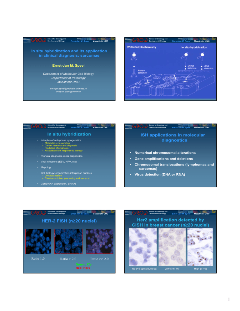
HER-2 FISH (n≥20 nuclei)
School for Oncology and
Developmental Biology
Ernst-Jan M. Speel
Maastricht UMC
Her2 amplification detected by
CISH in breast cancer (n≥20 nuclei)
•
Ratio 1.0
Ratio > 2.0
Ratio >> 2.0
Green: 17c
Red: Her2
No (<5 spots/nucleus)
Low (≥ 5 -9)
High (≥ 10)
1
School for Oncology and
Developmental Biology
Ernst-Jan M. Speel
School for Oncology and
Developmental Biology
Maastricht UMC
(Dis)advantages FISH vs CISH:
FISH
Maastricht UMC
Virus detection
CISH
Detection sensitivity
+
-
Multiple probes
+
-
Resolution
+
-
Nuclear overlap
±
±
Morphology
±
±
Preparation storage
±
+
Evaluation time
±
+
Out of focus
-
+
Autofluorescence
-
+
Special microscope
-
+
School for Oncology and
Developmental Biology
Ernst-Jan M. Speel
Ernst-Jan M. Speel
EBV RNA
“EBER”
Maastricht UMC
School for Oncology and
Developmental Biology
Ernst-Jan M. Speel
Maastricht UMC
School for Oncology and
Developmental Biology
Ernst-Jan M. Speel
Maastricht UMC
Essential steps in ISH procedure
Specimen preparation
- Accessibility probe/target
- Preservation morphology
Probe selection and labeling
Denaturation
(probe and cellular DNA)
In situ hybridization
Probe detection
Microscopy
Speel et al. Histochemistry 1999
School for Oncology and
Developmental Biology
Ernst-Jan M. Speel
Maastricht UMC
Preparation of biological specimen
• Solid support (glass, membrane) + coating
Optimal protocol for ISH on formalin-fixed,
paraffin-embedded tissue sections (1 day)
•
Deparaffination
•
85% formic acid / 0.3% H2O2 treatment, 5-20 min RT
•
1M sodium thiocyanate (NaSCN) treatment, 10 min 80oC
•
pepsin digestion, 4 mg/ml 0.02 M HCl 10-20 min 37oC
•
(Acid) dehydration steps
•
Post-fixation in 1% (para)formaldehyde, 10 min RT
•
ISH (denaturation 750C, hybridization 370C)
•
Stringent washings (2xSSC, 730C) and probe detection
• Fixation:
– Ethanol
– Methanol/Acetic acid
– (Para)formaldehyde
• Pretreatment:
– Proteolytic digestion (pepsin, proteinase K, other) or
microwave
– Detergents
– Endogenous enzyme inactivation
– RNase/DNase treatment
or 0.2 M HCl, 20 min RT
Hopman et al. Modern Pathol 4, 503-513, 1991; 1998; Vysis Inc
2
School for Oncology and
Developmental Biology
Ernst-Jan M. Speel
School for Oncology and
Developmental Biology
Maastricht UMC
Relation between nucleic acid target
and probe sequence
Ernst-Jan M. Speel
Maastricht UMC
ISH probes
• Locus-specific (single-copy)
• Repeat (centromere, telomere)
• Paint (chromosome (arm)
• Translocation
– Dual color, single fusion (2x flanking 1 side breakpoint)
– Extra signal (1x spanning breakpoint, 1x flanking)
– Dual color, dual fusion (2x spanning) breakpoint)
– Dual color, break apart (flanking 2 sides 1 breakpoint)
Speel, 1999
School for Oncology and
Developmental Biology
Ernst-Jan M. Speel
School for Oncology and
Developmental Biology
Maastricht UMC
Probe labeling and detection
Label ((d)NTPs)
Detection
Fluorochrome
Enzyme (HRP, APase)
Direct
Direct
Hapten
(bio, dig, FITC, DNP)
Indirect
(antibody and
avidine-biotin conjugates)
•
•
•
•
•
Temperature (37-420C)
pH (7.0)
Monovalent cations (Salt) (2xSSC)
Formamide (50-60%)
Probe
–
–
–
–
–
–
–
Chemical probe labeling: ULS etc.
Ernst-Jan M. Speel
Maastricht UMC
Fluorochromes often used in ISH
detection systems
Maastricht UMC
Parameters ISH
Enzymatic probe labeling: nick translation, random primed
labeling, PCR, endlabeling, tailing, in vitro transcription
School for Oncology and
Developmental Biology
Ernst-Jan M. Speel
DNA or RNA
Length
CG content
Base mismatches
Concentration
Dextran sulphate
Build-in haptens
School for Oncology and
Developmental Biology
Ernst-Jan M. Speel
Maastricht UMC
FISH: 1c / 7c copy number evaluation
Head and neck precursor lesions and
tumor resection margins
Disomy c1 (green)
Disomy c7 (red)
Trisomy c1 (green)
Polysomy c7 (red)
Bergshoeff et al., 2008
Definition CIN:
copy number differences between 1c and 7c, or polysomy
Now also Alexa, Spectrum, Platinum dyes, etc
3
School for Oncology and
Developmental Biology
Ernst-Jan M. Speel
School for Oncology and
Developmental Biology
Maastricht UMC
Ernst-Jan M. Speel
Maastricht UMC
Urine cytology:
bladder cancer cell detection
Spectral
Karyotyping
(SKY)
Normal
T(X,18)
Aneuploid
Lazar et al.
2006
(> 4 nuclei/preparation)
School for Oncology and
Developmental Biology
Ernst-Jan M. Speel
School for Oncology and
Developmental Biology
Maastricht UMC
Fine needle biopsy from retroperitoneum:
nonlipogenic area: desmoid tumor
Ernst-Jan M. Speel
Maastricht UMC
Conventional karyotyping: G-banding
• Advantages:
–
–
–
–
Global genetic information in single assay
Variants uncovered (undetectable by FISH and RT-PCR)
Diagnostically useful: fine needle biopsy, sensitive, specific
Provides direction for further molecular studies
• Limitations:
–
–
–
–
–
–
Ring chromosomes:
Chrom 12: green
MDM2: red
Requires fresh tissue
Mostly cell culture 1-10 days
Complex karyotypes, suboptimal morphology
False negatives due to cryptic rearrangements
Normal karyotypes (overgrowth normal fibroblasts, infiltrating cells)
Low cell density
Bridge, 2008
School for Oncology and
Developmental Biology
Ernst-Jan M. Speel
Maastricht UMC
ALT / WDL
School for Oncology and
Developmental Biology
n≥20 nuclei, different areas
Metaphase: MDM2
•
•
•
•
•
Accounts for up to 40-45% of all liposarcomas
Most prevalent in adults in the extremities and the retroperitoneum
Tendency to recur when occurring in deep anatomic sites
May dedifferentiate and metastasize in 2-20% of cases
Cytogenetics: Ring and giant marker chromosomes:
12q13-15 amplification: MDM2 + CDK4 candidate genes
Ernst-Jan M. Speel
FISH on Lipo(sarco)ma
Maastricht UMC
Red: CEP12
Green: MDM2 or CDK4
ALT / WDL: amplified MDM2 (l) and CDK4 (r)
ALT / WDL: amplified MDM2 with CEP12 alterations
Lipoma: 2 copies CDK4
4
School for Oncology and
Developmental Biology
Ernst-Jan M. Speel
Maastricht UMC
Conclusions: study on 50 lipo(sarco)mas
School for Oncology and
Developmental Biology
Ernst-Jan M. Speel
Maastricht UMC
Dual color, break-apart probes
FKHR/FOXO1
• Strong (2+) immunostaining of both MDM2 and CDK4 proteins show
a high amount of concordance with amplification of both gene loci in
ALT / WDL
• Lipomas show no amplification of MDM2 and CDK4 gene loci and
predominantly no protein expression
EWS
EWS
EWS
But:
• Weak (+) immunostaining of MDM2 correlates with the presence of
fat necrosis and inflammation in lipoma and ALT / WDL, but not with
amplification
• A subset of ALT / WDL shows no MDM2 and CDK4 amplification
EWS
Therefore:
• FISH might be preferred over IHC (and CDK4 over MDM2 IHC)
• What is genetic cause and implication of ALT without amplification?
FUS
CHOP
SYT
Bridge, 2008
School for Oncology and
Developmental Biology
Ernst-Jan M. Speel
Maastricht UMC
Dual color,
Break-apart
EWS
FISH probe
School for Oncology and
Developmental Biology
Ernst-Jan M. Speel
Maastricht UMC
Synovial sarcoma
• It accounts for up to 5-10% of all soft tissue
sarcomas
• 5 years survival 36-76%
• Most prevalent in adolescent and young
adults in deep soft tissue of the lower
extremities
• Specific t(X,18) translocation is found in 90%
of synovial sarcoma
• Genes affected by the t(X,18) are SYT from
chromosome 18 and SSX1, SSX2 and
SSX4 from the X chromosome
Lazar et al. 2006
School for Oncology and
Developmental Biology
Ernst-Jan M. Speel
Maastricht UMC
School for Oncology and
Developmental Biology
Ernst-Jan M. Speel
Maastricht UMC
T(X,18) FISH
Vysis, Abbott Molecular
Evaluatie:
n≥50 nuclei
n≥50 nuclei
A+B:
Geen translocatie
C:
Translocatie
D-H:
Kans op translocatie
No translocation
Translocation
5
School for Oncology and
Developmental Biology
Ernst-Jan M. Speel
Maastricht UMC
Molecular cytogenetics: FISH
School for Oncology and
Developmental Biology
Ernst-Jan M. Speel
Maastricht UMC
Tyramide signal amplification
• Advantages:
–
–
–
–
–
–
–
–
Fresh, frozen, paraffin-embedded material (interphase and metaphase)
Localize alteration (translocation) in specific cells and tissue types
Useful if tumor is heterogeneous, or in case of MRD
Diagnostically useful: fine needle biopsy, sensitive, specific
Can provide results if karyotyping or RT-PCR is inconclusive
Rapid turn-around time
Validation and implementation easy
Normal tissue parts can serve as FISH control
• Limitations:
HPV
– Targeted approach, except for CGH and SKY analysis
– Relatively gross approach (no information on fusion genes and variants)
– Number of commercially available probes is limited (Abbott Molecular,
Kreatech)
– Requires fluorescence microcope
– Interpretation may be challenging, expertise required
– Period of storage
School for Oncology and
Developmental Biology
Ernst-Jan M. Speel
Maastricht UMC
Speel et al., 1997; 1998; Hafkamp et al., 2003; 2008
School for Oncology and
Developmental Biology
Ernst-Jan M. Speel
Acknowledgements
Good luck
with
evaluation!
Maastricht
Molecular Cell Biology
Ton Hopman
Sandra Claessen
Annick Haesevoets
Frans Ramaekers
Pathology
Els Meulemans
Andrea Ruland
Guido Roemen
David Creytens
Clinical Genetics
Josefa Albrechts
Jannie Janssen
Merryn MacVille
Epidemiology
Adri Voogd
Maastricht UMC
Maastricht
Otorhinolaryngology &
Head and Neck Surgery
Ewa Bergshoeff
Harriet Hafkamp
Hans Manni
Bernd Kremer
Nijmegen
Pathology
Piet Slootweg
Rotterdam
Pathology
Winand Dinjens
Gent
Pathology
Patrick Pauwels
6
